
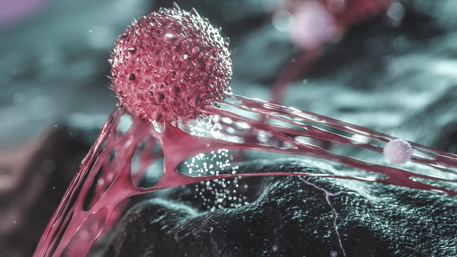
A common sugar found in everyday foods and sweetened beverages may play a surprisingly critical and unexpected role in helping aggressive ovarian cancer spread throughout the body. Researchers at The Wistar Institute have made a groundbreaking discovery showing that cancer cells which manage to survive chemotherapy can produce and release fructose. Instead of merely acting as an energy source, this secreted sugar acts as a potent biological signal, encouraging nearby tumor cells to break free from their original locations and initiate deadly metastasis.
The findings, recently published in the prestigious scientific journal Nature Aging, reveal a previously unknown mechanism through which cancer cells can continue to actively influence and worsen a tumor’s microenvironment even after traditional medical treatments have successfully stopped them from multiplying. Beyond illuminating the complex biology of tumor survival, this discovery raises profound and urgent questions about whether dietary sugar intake and commonly prescribed cholesterol-lowering medications might inadvertently affect how cancer progresses in patients.
"Some cancer cells that survive chemotherapy aren’t dividing anymore, but they’re still biologically active," explained Aidan Cole, Ph.D., a postdoctoral fellow in the laboratory of Katherine Aird, Ph.D., at The Wistar Institute and the first author on the new study. "Instead, they continue to release molecules that send signals to nearby cells. Our study is among the first to show that a nutrient—in this case, fructose—can act as one of those signals."
Why Ovarian Cancer Often Returns After Chemotherapy
To fully grasp the significance of these findings, medical researchers must contend with one of the most persistent and devastating challenges in modern oncology: the high recurrence rate of ovarian cancer. The vast majority of patients diagnosed with ovarian cancer receive platinum-based chemotherapy as a standard course of treatment. These powerful pharmaceutical drugs are specifically designed to infiltrate and damage the DNA of cancer cells, thereby preventing them from reproducing and causing the tumor to shrink.
Although many patients initially respond exceptionally well to these rigorous therapies, the cancer eventually returns in the vast majority of cases. Furthermore, when the disease recurs, it frequently does so in a disseminated pattern, spreading extensively throughout the abdominal cavity. This anatomical space houses vital organs such as the stomach, intestines, and liver, making subsequent treatments exceptionally difficult.
This dangerous spread, formally known as metastasis, is directly responsible for approximately 90 percent of all ovarian cancer-related deaths. Understanding the precise cellular and molecular mechanics that allow cancer cells to escape from their primary tumor site, invade surrounding tissues, and establish successful secondary colonies is therefore a major, high-priority focus for researchers worldwide.
Previous scientific studies have long suggested that a subset of cancer cells surviving chemotherapy can actively contribute to disease recurrence, even when those specific cells are no longer actively dividing or multiplying. These resilient surviving cells can persist within the body and continue producing and releasing a complex mixture of biochemical substances that profoundly influence the behavior, growth patterns, and aggressiveness of surrounding cells.
To investigate this elusive process in meticulous detail, Cole and his multidisciplinary team of colleagues developed an innovative experimental model. They effectively separated the surviving cancer cells from the various molecules they secreted into their environment. Through this careful isolation, the researchers made a striking discovery: the released substances alone, completely independent of the physical presence of the surviving cells themselves, were sufficient to dramatically increase cancer spread in a preclinical model.
"As far as we know, this is the first time anyone has shown, in a preclinical model rather than just a dish, that it’s the molecules these cells release—not the cells themselves—that drive the cancer’s spread," Cole noted.
A Common Sugar Emerges as a Surprising Cancer Signal
Once the team established that secreted molecules were driving the aggressive behavior, the next major challenge was identifying which specific substances were responsible for encouraging neighboring cancer cells to break free and migrate. Through rigorous analysis, the researchers uncovered an unexpected and ubiquitous culprit: fructose.
Fructose is a naturally occurring simple sugar found abundantly in fruit, honey, and a wide variety of whole foods. However, it is also a primary component of table sugar and high-fructose corn syrup, an industrial sweetener that is heavily utilized in commercial soft drinks, baked goods, and an extensive array of ultra-processed foods consumed daily by millions of people around the globe.
The study revealed that cancer cells surviving chemotherapy possess the ability to synthesize fructose internally and actively release it into their immediate extracellular surroundings. Rather than simply serving as metabolic fuel or a basic energy source—which is the traditional role attributed to sugars in cellular biology—this particular fructose acted as an active chemical messenger. It signaled neighboring cancer cells, altering their internal programming and making them significantly more capable of migration and metastasis.
Equally concerning, the researchers uncovered another alarming possibility during their laboratory investigations. In preclinical experiments, exposing cells to high amounts of fructose—concentrations directly comparable to those consumed by humans who regularly drink sugary beverages—could promote the very same biological signals associated with cancer spread, even in the complete absence of prior chemotherapy treatment.
This compelling observation strongly suggests that everyday human diet might directly influence cancer progression through a biochemical mechanism that scientists had not previously recognized or understood. Fructose consumption is an especially relevant public health consideration in the United States, where high-fructose corn syrup can account for approximately 8 percent to 20 percent of total daily calorie intake for certain individuals.
Unlike many genetic, environmental, or demographic factors that influence overall cancer risk and disease progression, dietary fructose intake is a modifiable behavior. This reality raises the intriguing possibility that targeted nutritional strategies and dietary modifications might eventually become integrated into comprehensive clinical efforts to limit cancer spread and improve patient prognoses.
However, researchers issue a firm note of caution, emphasizing that scientific teams have not yet formally tested whether reducing dietary fructose consumption actually improves clinical outcomes in human patients diagnosed with ovarian cancer. The current findings do not definitively establish that eating sugar directly causes cancer to spread in humans, and a significant amount of additional clinical research is urgently required before any specific, evidence-based dietary recommendations can be responsibly formulated on the basis of this newly discovered mechanism.
How Fructose Helps Cancer Cells Break Free
To understand the exact cellular mechanics of how fructose encourages cancer spread, the research team conducted a deep dive into the biochemical changes occurring inside cancer cells when they are exposed to the sugar.
To accomplish this, they utilized a suite of advanced molecular biology techniques, most notably a CRISPR screen. This cutting-edge technology allows modern scientists to systematically examine and disrupt thousands of genes simultaneously, making it possible to pinpoint precisely which genetic pathways influence a specific, complex cellular process.
The results of these intricate experiments revealed a fascinating and unexpected chain of events: exposure to fructose actively suppresses cholesterol production in neighboring cancer cells.
Although cholesterol is most frequently discussed in popular culture and public health messaging in connection with cardiovascular disease, clogged arteries, and heart attacks, it also serves a multitude of vital, indispensable functions throughout the human body. Beyond its systemic roles, cholesterol is an essential structural component of cell membranes—the delicate outer lipid layers that completely surround every cell and help strictly regulate how those cells interact with their external environment, neighboring cells, and chemical signals.
In the specific context of epithelial and tumor tissues, cellular cholesterol helps maintain the physical connections and adhesion molecules that tightly hold neighboring cells together. These specialized structures act somewhat like a biological glue, creating a cohesive tissue architecture that makes it structurally difficult for individual cancer cells to separate from one another and migrate away.
When fructose downregulates and reduces internal cholesterol production within the cancer cells, those crucial cellular connections weaken significantly. Lacking their usual structural cohesion, the cancer cells can detach from the primary tumor mass much more easily. This loss of adhesion potentially allows them to escape their localized confines, enter surrounding tissues, and spread successfully to distant anatomical locations.
This remarkable discovery provides a coherent, mechanistic explanation for how a common nutritional component can directly influence one of the most lethal and elusive aspects of cancer biology.
A Surprising Connection Between Statins and Cancer Spread
The unexpected finding regarding cholesterol suppression immediately led the investigative team to pursue another vital and potentially impactful line of inquiry: exploring whether widely prescribed cholesterol-lowering medications, known as statins, might share a similar biological effect.
Statins are among the most frequently prescribed drugs in the world, with approximately 39 million people in the United States alone taking them regularly. These pharmaceutical agents work primarily by inhibiting an essential enzyme in the liver, thereby reducing the body’s overall production of cholesterol to lower the ongoing risk of heart attacks, strokes, and cardiovascular morbidity.
Because the research demonstrated that fructose promoted cancer cell migration specifically by suppressing cholesterol production, the scientific team needed to determine whether taking statins—which also drastically lower cholesterol levels—might inadvertently produce similar pro-metastatic effects in cancer patients.
Preliminary laboratory experiments revealed that exposing cancer cells to statins alone could indeed weaken the adhesive connections between cells, making it substantially easier for them to separate, detach, and escape their tissue matrix.
This provocative finding immediately raises complex clinical questions about whether commonly used cholesterol-lowering medications could potentially influence the efficacy of chemotherapy or alter metastatic trajectories in certain patient populations. Researchers are actively pursuing follow-up studies to investigate this possibility thoroughly, but they emphasize strongly that these laboratory results have not yet been demonstrated in human patients. Consequently, the findings do not constitute any reason whatsoever for patients to discontinue taking their prescribed statins, which remain a life-saving intervention for cardiovascular health.
"We haven’t tested this effect in patients yet, but it raises questions about combining cholesterol-lowering drugs with chemotherapy, especially since ovarian cancer is most common in postmenopausal women who are often already on statins," said Katherine Aird, Ph.D., professor and co-leader of the Molecular and Cellular Oncogenesis Program in the Ellen and Ronald Caplan Cancer Center at The Wistar Institute, and senior author of the study.
Could Fructose Also Help Other Cancers Spread?
Although the primary scope of this newly published research focused explicitly on ovarian cancer, the investigators strongly suspect that the exact same underlying biological processes and signaling pathways could be at play in several other forms of the disease.
Many aggressive cancers that originate and affect organs located deep within the human torso and abdominal cavity—such as pancreatic, colorectal, and liver cancers—share similar microenvironments and are likewise notorious for invading nearby tissues and spreading extensively throughout the body. If dietary or metabolically produced fructose influences cellular adhesion and cholesterol production in these other malignancies in a comparable manner, the broad implications of this discovery could extend far beyond the field of gynecological oncology.
"We think other cancers that spread within the torso—pancreatic, colon, liver—could behave similarly. We can’t call it universal yet, but we think the effects are not just limited to ovarian cancer," Aird stated.
Demonstrating this broader applicability is already underway. Aird, Cole, and their laboratory colleagues have initiated the design and execution of subsequent experimental phases to determine whether these striking findings can be successfully reproduced in pancreatic, colon, and liver cancer models.
Ultimately, this research opens an entirely new chapter in how scientists understand the complex dynamics of metastasis, particularly in the difficult aftermath of chemotherapy. It also serves as a potent reminder that the intricate interplay between everyday nutrients, internal metabolic pathways like cholesterol synthesis, and cancer cell behavior is vastly more complicated than previously understood.
While definitive answers regarding the ultimate clinical implications for diet and pharmacological interventions remain on the horizon, identifying fructose as a biochemical signal that helps chemotherapy-surviving cancer cells break free marks a significant step forward. In time, these insights could guide researchers toward innovative, targeted strategies for preventing metastasis and fundamentally improving patient treatment outcomes.