
Why do some new skills seem to click almost instantly in the human mind while others remain frustratingly difficult to master, even after hours of repeated practice and immense personal effort? According to a team of neuroscientists, the difference may involve far more than innate talent, individual aptitude, or sheer willpower. It may fundamentally depend on whether the physical brain is in the right physiological condition to successfully turn temporary practice into lasting, permanent learning.
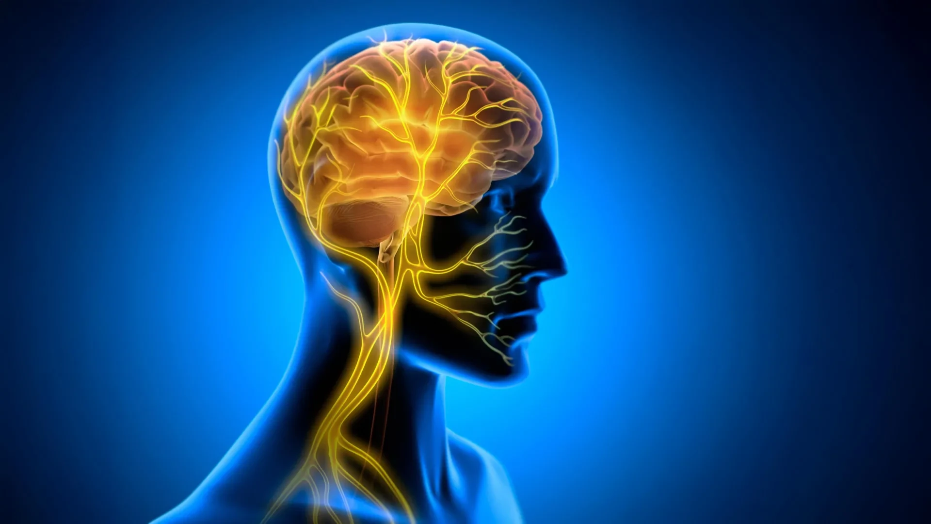
For generations, the scientific study of learning has largely treated the brain as an isolated command center, focusing intensely on neural circuits, synaptic connections, and cognitive processing while largely ignoring the rest of the physical body. However, a growing body of contemporary research demonstrates that learning does not happen independently of the rest of the body. Internal organs are constantly sending a vast stream of physiological information directly to the brain through the vagus nerve, which serves as one of the central nervous system’s major communication pathways.
Addressing this complex brain-body interaction, researchers at Tohoku University who specialize in super-network brain physiology have now shown in a groundbreaking study involving mice that artificially stimulating this crucial nerve immediately after training can significantly strengthen long-term motor learning. Their experimental findings point directly to a previously overlooked role for continuous communication from the body to the brain, revealing how somatic signals actively help new physical skills persist over time.
The comprehensive findings from this research were officially published in the scientific journal iScience on August 25, 2026, opening up entirely new avenues of inquiry into how humans and animals acquire and consolidate new abilities.
The Vagus Nerve and Learning
To understand the significance of the Tohoku University study, one must look closely at the vagus nerve itself. Serving as a major information highway running continuously between the brain and the body, the vagus nerve carries a dense volume of vital signals upward from internal organs such as the heart, lungs, and digestive tract to the brainstem. Simultaneously, it transmits crucial motor and regulatory instructions downward from the brain back to those very same organs, maintaining a delicate, continuous feedback loop.
Scientists have long known that they can artificially influence this vital communication pathway using a medical technique known as vagus nerve stimulation (VNS). Vagus nerve stimulation is already clinically approved and widely utilized in modern medicine for treating several challenging medical disorders, including drug-resistant epilepsy and severe forms of depression.
In earlier academic research, scientists have primarily explored VNS through the lens of neuromodulation. Under this traditional view, stimulation alters electrical activity and chemical signaling within various neurotransmitter systems in the brain, such as norepinephrine and acetylcholine pathways, which are known to modulate attention and plasticity. However, the new study from Tohoku University suggests that there may be another profoundly important mechanism involved in how VNS aids recovery and skill acquisition: dynamic, rhythmic changes in blood vessels located deep inside the brain tissue itself.
To investigate this compelling possibility, the research team designed a sophisticated experimental setup. They created a small, specialized cuff electrode engineered to remain safely and continuously attached to the left cervical vagus nerve in laboratory mice. With this hardware in place, the scientists tested the effects of VNS during horizontal optokinetic response (HOKR) learning.
The HOKR is a well-established, cerebellum-dependent eye movement task that trains mice to systematically improve their visual-tracking ability, specifically enhancing their capacity to smoothly follow moving visual stripes with their eyes. This natural biological response is remarkably similar to the automatic, compensatory eye movements that a human passenger makes while standing still on a station platform and watching a high-speed train move past their field of vision. By monitoring this precise motor task, the researchers could accurately measure how well the animals were learning and retaining the visual-tracking skill over multiple days.
The Biggest Effect Appeared After Practice
One of the most surprising and revealing aspects of the new study involves the precise timing of the intervention. Rather than applying vagus nerve stimulation continuously during the learning task itself, the researchers chose to apply VNS strictly after each daily training session had concluded.
Interestingly, the artificial stimulation did not produce an immediate, miraculous improvement in performance while the mice were actively engaged in the training task. The animals did not suddenly track the moving stripes better mid-exercise simply because the nerve was being stimulated at that exact second. Instead, the profound benefits of the stimulation became apparent much later.
On the days following the training sessions, the mice that had received the post-training vagus nerve stimulation demonstrated significantly stronger, more robust long-term learning compared to control groups. This clear, consistent performance pattern strongly suggests that the nerve stimulation actively influences biological processes that happen after active practice has ceased—a critical window of time when the resting brain is busy consolidating what it has just learned, transforming fragile short-term experiences into more durable, long-lasting memory traces.
"The key point is that VNS was delivered only after training," explains Professor Ko Matsui, a senior researcher involved in the study. "Our findings suggest that VNS may open a hidden window of opportunity for enhanced learning by making the brain environment more receptive to long-lasting change."
Rhythmic Changes in Brain Blood Volume
Intrigued by the delayed nature of the memory enhancement, the research team set out to look deeper inside the brain for physiological changes that might directly accompany the improved learning outcomes. Specifically, they decided to measure local blood volume activity in the neural tissue near the cerebellar flocculus, a distinct anatomical area of the brain that is heavily involved in mediating HOKR learning and motor coordination.
Utilizing an advanced optical technique known as fiber photometry, the researchers made a striking discovery. They observed that a single round of vagus nerve stimulation produced a distinct, two-phase vascular response within the targeted brain region. First, the local blood volume briefly decreased, which was followed immediately by a pronounced rise after a slight physiological delay.
More importantly, when the vagus nerve stimulation was systematically repeated, it created sustained, rhythmic oscillations in the volume of blood flowing through the brain’s microvasculature. These rhythmic vascular movements were not random physiological noise; rather, they appeared to be deeply intertwined with the learning process itself.
When the researchers analyzed individual animals, they found that mice exhibiting larger blood volume oscillations generally demonstrated significantly better learning performance by the fifth day of the experiment. This strong correlation suggests that dynamic shifts in the brain’s internal vascular environment are directly connected to the long-lasting neuroplastic effects of vagus nerve stimulation, providing a physical mechanism for how body-to-brain signaling supports cognitive persistence.
"Our brains may be more strongly influenced by the body than we imagine," says lead author Junyu Chen, elaborating on the implications of the team’s data. "By tuning the brain’s metabolic environment, including rhythmic vascular movements, we may eventually unlock capacities that would otherwise remain latent."
Exploring the Brain-Body Connection
As the scientific community digests these findings, researchers are already looking ahead to the next phases of investigation. Future research efforts will focus heavily on refining vagus nerve stimulation protocols and determining with greater precision the exact cellular and molecular mechanisms by which communication between the body and the brain supports long-term neural plasticity.
By studying this complex, two-way communication pathway in much greater detail, scientists hope to build a comprehensive framework for understanding how transient daily experiences are successfully converted into permanent knowledge. Ultimately, this line of inquiry may illuminate how these foundational neurobiological processes can eventually be safely enhanced in clinical and educational settings, bridging the gap between physical somatic health and cognitive mastery.